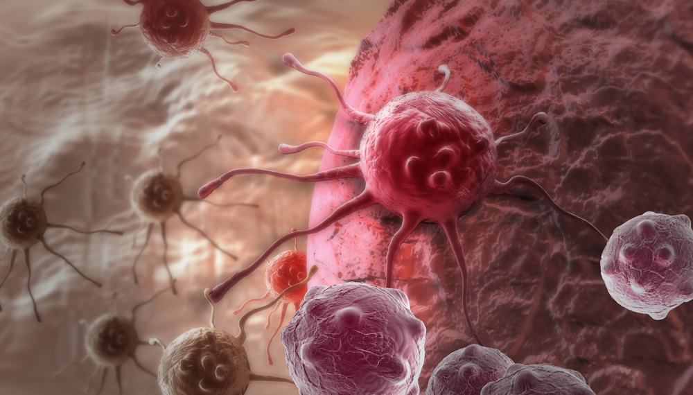
Tumor markers (TM) refer to the synthesis and secretion of tumor cells and gene expression during the occurrence and proliferation of malignant tumors, or the abnormal production and elevation of the body’s response to tumors. Tumors exist and grow. a class of substances.
A tumor antigen can be a tumor marker, but a tumor marker is not necessarily a tumor antigen. TM exists in the blood, body fluids, cells or tissues of patients and can be determined by biochemical, immunological and molecular biology methods, and has certain value in the auxiliary diagnosis, differential diagnosis, curative effect observation, detection of recurrence and prognosis evaluation of tumors. .
The classification and nomenclature of tumor markers have not been completely unified, and they can generally be divided into embryonic antigens, sugar chain antigens, enzymes, hormones, proteins and gene markers.
I. Common tumor markers:
1. Embryonic antigenic tumor markers
Embryonic antigens are normal components produced by embryonic tissue in the embryonic development stage. They decrease in the late embryonic stage and gradually disappear or only exist in very small amounts after birth. These embryonic antigens reappear in cancer patients.
Includes:
-Alpha-fetoprotein
-Carcinoembryonic antigen
2. Carbohydrate antigen tumor markers
Carbohydrate antigens (CAs) are antigens formed due to abnormal glycosyl residues in cell membrane glycoproteins. “CA” also means tumor, formerly known as cancer antigen. There are abundant glycoproteins on the surface of normal cell membranes. When normal cells are transformed into malignant cells, the glycoproteins on the cell surface mutate to form a special antigen different from normal cells, which usually exists on the surface of tumor cells or is secreted by tumor cells. .
Includes:
-CA125
-CA19-9
-CA15-3
3. Enzymatic tumor markers
Most enzymes are present in cells, which are released into the blood due to the destruction of tumor cells or changes in cell membrane permeability; abnormal metabolism of tumor cells increases the synthesis of certain enzymes or isozymes; compression and infiltration of tumor tissue lead to The excretion of certain enzymes is blocked, resulting in abnormally elevated enzyme activity in the serum of tumor patients.
Includes:
– Neuron-specific enolase
-α-L-fucosidase
4. Hormonal tumor markers
Certain tissues that do not produce hormones under normal circumstances can produce and release some peptide hormones (ectopic endocrine hormones) and lead to corresponding syndromes during malignant transformation. Therefore, the elevation of these endocrine hormones can also be used as tumor-related markers .
Includes:
– Human chorionic gonadotropin
-Catecholamines
5. Protein tumor markers
Most solid tumors are derived from epithelial cells. When tumor cells differentiate and proliferate rapidly, some cell types or components that do not appear in normal tissues, such as keratin as a cytoskeleton, become tumor markers.
Includes:
-Prostate specific antigen
– Cytokeratin 19
-Ferritin
Common tumor markers in physical examination
Common physical examination items can be divided into the following categories:
– Serum carcinoembryonic antigen (CEA): normal value less than or equal to 3.45 micrograms/liter. Initially found to be elevated in colon cancer patients, CEA was later found to be elevated in 30% of patients with gastric, urethral, ovarian, lung, pancreatic, breast, medullary thyroid, bladder and cervical cancers. Elevated CEA.
– Alpha-fetoprotein (AFP): AFP is the earliest tumor marker discovered, and it is a common test item for the diagnosis of primary liver cancer. About 87% of primary liver cancer patients have AFP as high as 20 micrograms/liter or more.
-Prostate-specific antigen (PSA): the normal value is less than 4 micrograms/liter, the positive rate in prostate cancer is as high as 30% to 86%, and its elevated level is closely related to the tumor.
– Chorionic Gonadotropin (HCG): The concentration of normal human blood is less than 5 micrograms/L, such as those with chorioepithelial carcinoma, embryonal malignant teratoma of testis and ovary, HCG can be elevated, and blood and urine are The amount of HCG is associated with prognosis.
use:
– Early detection of tumors;
-Tumor screening and screening;
– diagnosis, differential diagnosis and staging of tumors;
-Monitoring the curative effect of surgery, chemotherapy and radiotherapy in tumor patients;
– indicators of tumor recurrence;
– Prognostic judgment of tumor;
– Find the primary tumor of metastatic tumors of unknown origin.
2. Selection of tumor markers Combined application of multiple markers
Recommendations for the application of tumor markers:
– Patients after surgery should have tumor markers measured every 2-3 months for at least 2 years. In the absence of treatment, at least 2 consecutive (two time intervals) tumor markers showed a linear increase, which can be considered as tumor recurrence.
-In patients under treatment, the increase of tumor markers means the deterioration of the disease (meaning that the measured value of tumor markers increases by 25% as deterioration). Check again every 2-4 weeks for reliability.
– There are too many tumor markers, and the sensitivity or specificity of a single marker is often low, which cannot meet the clinical requirements. In theory and practice, simultaneous determination of multiple markers is recommended to improve sensitivity and specificity.
– Tumor markers are not the only basis for tumor diagnosis, and clinical symptoms, imaging examinations and other methods should be considered comprehensively. The diagnosis of tumor must be based on histological or cytopathological diagnosis.
– Due to individual differences in patients, specific clinical conditions of patients and other factors, the analysis of tumor markers must be combined with clinical conditions and compared from multiple perspectives in order to draw objective and true conclusions.
– Certain tumor markers can also be abnormally elevated under certain physiological conditions or in certain benign diseases, and attention should be paid to identification.


