Breast cancer is one of the most common malignant tumors in women. In many countries, breast cancer ranks first in the mortality rate of female malignant tumors. Breast cancer recurrence and metastasis are the main causes of death in breast cancer patients. About 24% to 30% of patients with negative lymph nodes have recurrence and metastasis, and the rate of recurrence and metastasis in patients with positive lymph nodes is as high as 50% to 60%. The 5-year survival rate for cancer is only 26%.
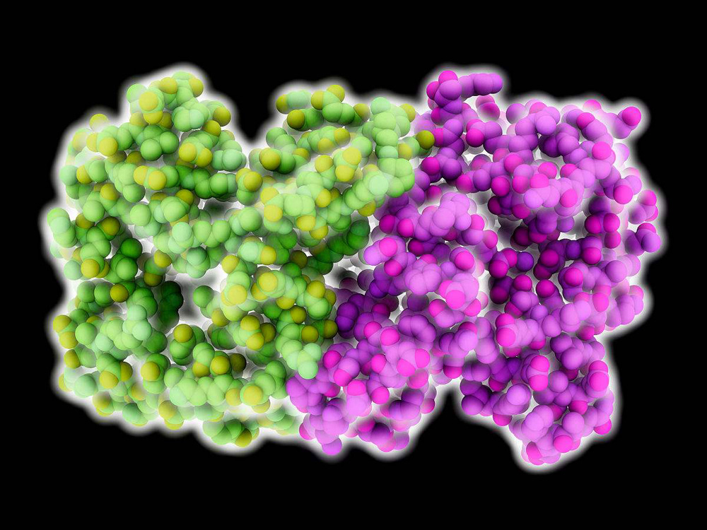
Firefly luciferase is a commonly used signaling molecule that can be used to label tumor cells.
One. cell method
The eukaryotic expression recombinant plasmid pRc/CMV2-7 with the luciferase luc-transferase reporter gene was screened by G418 to obtain a stable human breast cancer cell line MCF-7-luc, and the luciferase gene was stably expressed in cells. Clone MCF-7 was evaluated for its luminescence ability in vitro. By establishing a nude mouse xenograft model, and using the in vivo bioluminescence imaging system to detect the tumor growth and metastasis, it provides an ideal situation for the next step to monitor the changes of the nude mice xenografts on the drug effect and to analyze the drug’s therapeutic effect on the tumor.
Determination of G418 Screening Concentration:
The cells were cultured in a cell incubator at 37℃, and six concentration gradients of G418 were set at 0, 200, 400, 600, 800, and 1000 μg/ml. 24h after the cells were seeded, they were added to a 6-well plate, and 2 wells of each concentration were set to die within 10-14 days. The cell growth was observed every day, and the G418 concentration with the lowest death was the concentration of screening plasmid-transfected cloned MCF-7 cells.
1) Cell transfection
Take MCF-7 cells in logarithmic growth phase and inoculate the cells in a 6-well plate until the cell confluence reaches 80% to 90% of the well plate, and then TM can be transfected. Transfection was performed according to the instructions of Lipofectamine2000 kit. Add luc plasmid pRc/CMV2 at 8 μg/ml to 250 μl of serum-free, double-antibody-free DMEM medium. Add 10 μl LipofectamineTM2000 to 250 μl of serum-free, double-antibody-free DMEM medium, and incubate at room temperature for 5 min. Mix the above two dilutions, incubate at room temperature for 30 min, add them to the cell well plate, and place them in an incubator for conventional culture.
Monoclonal Cell Screening
24h after transfection, the cells were trypsinized and seeded into a new 6-well plate at a ratio of 1:6, and the experimentally determined concentration of G418 was added at the same time, and then the medium was changed every 2d and the G418 selection was maintained until the emergence of single-cell resistant clones. Single-resistant clones were selected to 96-well plates, and then transferred to 24-well plates to continue subculture after they gradually proliferated.
3) Luciferase activity identified positive clones
The luciferase activity was detected by LuciferaseAs-sayAystem when the single resistant clones were passaged to the fifth passage. During detection, each clone was seeded into a 24-well plate at 1×10⁵ cells/well. After 24 hours, the cell lysate was lysed at 12,000 rpm and centrifuged at 4°C for 10 min. The lysate was collected, and 10 μl of the supernatant was added to a 96-well white plate, and 50 μl was added to each well. The luciferase 96 microplate luminometer reads the substrate continuously. After 2 s, the fluorescence value (RLU) for 10 s is taken. Each clone has 3 duplicate wells, and the cell clones with high RLU value are kept for subculture. After another 5 generations, luciferin Enzyme activity assay. The clones with higher RLU values were retained until the 30th passage, and the clones with the highest luciferase activity, MCF-7-luc, were positive clones.
Fluorescence values of different numbers of cell clones detected 7-luc positive clones. The screened MCF-4×104,2×104,1×104,5000,2500,1250,625,312 and 156 were inoculated into In the 96-well black plate, the other group had only cells, and the other group had only medium as a control. Two duplicate wells were set up. The supernatant was routinely cultured and washed twice with PBS. The concentration was 150 μg/ml, PBS, and D- was added immediately to detect the correlation between the luminescence intensity and the number of cells by an in vivo imaging system.
4) Drawing of cell growth curve
MCF7-luc cells and MCF-7 cells expressing luciferase were taken as a control and seeded in a 24-well plate at a seeding density of 2×10⁴/well. 1-7 days after cell inoculation, 3 wells of cells were digested with trypsin every day, and the number of cells was measured with a cell counter. Taking the days of cell growth as the abscissa and the number of cells as the ordinate, two cell growth curves were drawn respectively.
Two. animal model
Establishment of BLAB/c Nude Mice Subcutaneous Tumor Model BLAB/cnu/nu nude mice, 4 to 5 weeks old, body weight (15±2) g, 3 males and 3 females. MCF-7 in logarithmic growth phase was resuspended in PBS to 2.5×10/ml suspension, and 100μl was inoculated subcutaneously on the left and right dorsal sides of each nude mouse near the axilla, for a total of 6 mice. On the 5th day after inoculation, the in vivo imaging system of German company BERTHOLD was used to detect the signal intensity. After every 5d observation, a continuous observation for 30d. Before observation, each nude mouse was anesthetized with sodium pentobarbital (measurement: 35 mg/kg body weight), intraperitoneally injected with luciferin (invivograde) at a dose of 150 mg/kg body weight, and 10 minutes later, in vivo imaging was performed to observe the growth of subcutaneous tumors. Fluorescence values at each time point were analyzed. Plot the tumor subcutaneous growth curve.
Pathomorphological observation of subcutaneous transplanted tumor of MCF-7-luc cells in nude mice
Twenty-five days after the subcutaneous inoculation of MCF-7-luc cells in nude mice, the mice were sacrificed by cervical dislocation. The tumor tissue was taken and made into paraffin sections with a thickness of 3 μm. The pathological morphology of the cells was observed after HE staining.
Live animal imaging technology is a new type of stable and reliable developed recently, and it is a powerful method for detecting molecular and cellular events in animals. Bioluminescence imaging (BLI) can be used to visually and accurately detect the size of living lesions without damage.


